
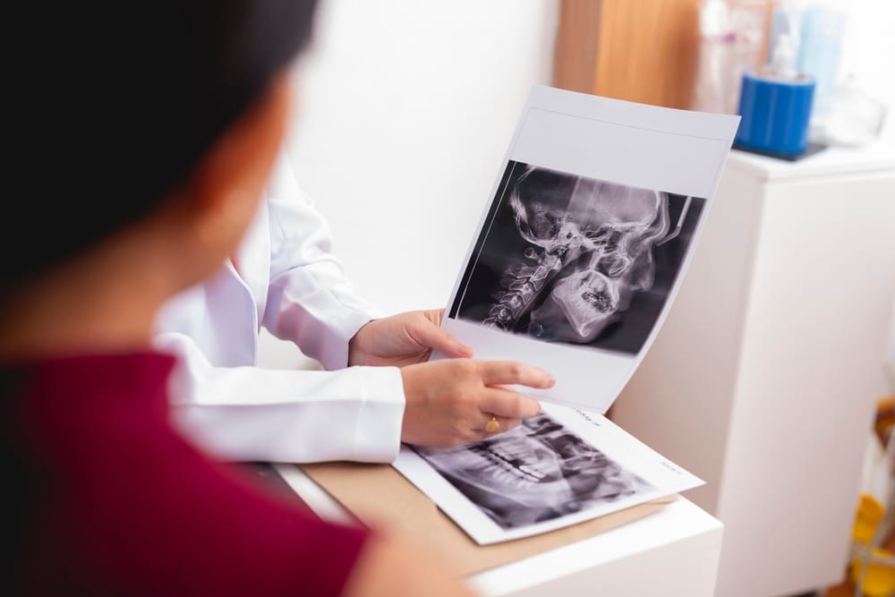
Bone grafting for dental implants often bridges the gap when there isn’t enough jawbone for a stable implant. Guided bone regeneration, or GBR, is the specific procedure that adds that missing bone back.
It uses a barrier membrane to protect the area while new bone grows underneath. Over time, that slow growth builds the volume an implant needs to stay stable for years. For many patients, this single step is what turns “not a candidate” into a real treatment plan.
Key Takeaways
- Barrier membranes block soft tissue and epithelial cells from entering the defect site, creating the protected space where osteogenic cells and a stable fibrin clot can build new lamellar bone.
- Graft materials range from autogenous bone taken from the patient’s own body to composite grafts enhanced with platelet-rich fibrin or recombinant human bone morphogenetic protein-2.
- The dental team selects either a simultaneous or staged approach based on bone loss severity, and CBCT imaging maps the defect before any treatment plan is finalized.
- GBR is a well-documented and predictable procedure, and speaking with a dental professional is the right next step if you think you may be a candidate.
- Common patient questions about healing, oral hygiene, and what to expect after the procedure are answered in the FAQs section below.
Barrier Membranes and the Role of Soft Tissue Exclusion in Guided Tissue Regeneration
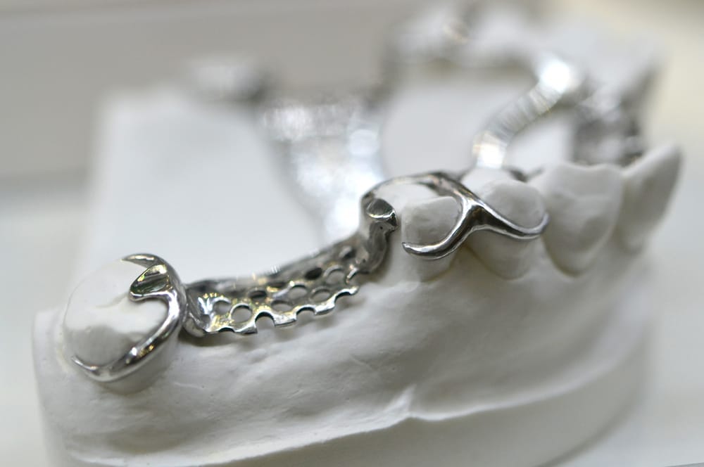
Barrier membranes are the defining feature of guided bone regeneration. They exist to solve one specific problem inside the mouth: soft tissue grows faster than bone.
When bone loss comes from periodontal disease or gum disease, the open defect fills with soft tissue fast. Epithelial cells move in before slower-growing bone cells get a chance to settle. A membrane over that defect blocks the invasion and creates a protected pocket where new bone can actually form.
Without that barrier, the body simply repairs the gap with soft tissue instead of solid bone. That soft tissue won’t support an implant long term.
Resorbable vs. Non-Resorbable Membranes
The two membrane types mainly differ in how dentists remove them once healing wraps up.
Resorbable membranes, most often collagen membranes, break down naturally inside the body. Removing them doesn’t require a second surgery, which makes them a common choice for moderate bone deficiency cases.
Non-resorbable membranes, including PTFE membranes and titanium mesh, hold their shape better under pressure. Titanium mesh works well for vertical ridge augmentation because it resists collapse when soft tissue pushes against it. The trade-off is a follow-up visit to remove the membrane once healing finishes.
The PASS Principle
Dentists use the PASS principle as a checklist for predictable outcomes. The four conditions are:
- Primary wound closure
- Angiogenesis, or new blood vessel growth
- Space creation
- Blood clot stability
Getting all four right takes practice, and skipping one often explains why a graft underperforms. Gum pockets or compromised gingival tissue make membrane coverage one of the toughest parts of the whole procedure.
Bone loss from periodontal disease or gum disease is often what brings patients to this point in the first place. GBR doesn’t treat the underlying disease, but it does rebuild the volume that disease has eroded. Oral health must be stabilized before bone augmentation can move forward.
Graft Material Options: From Autogenous Bone Grafts to Composite Graft Advances
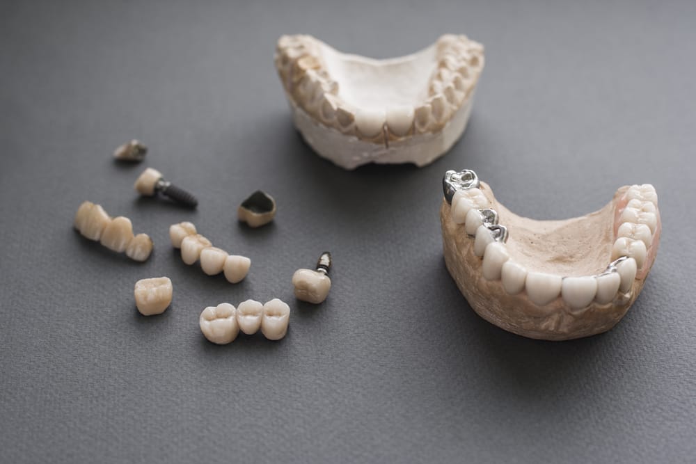
The material beneath a membrane needs to support new bone growth actively. The right choice depends on the defect size and the patient’s health history. It also depends on what the dental team expects will work best at that site. No single material fits every case, which is why several options exist below.
Autogenous Bone: The Gold Standard
Autogenous bone grafts use bone from the patient’s own body. This material carries living osteogenic cells directly into the graft site, including mesenchymal stem cells, osteoprogenitor cells, and pluripotential cells. These cells can turn into new bone tissue over time.
Autogenous bone chips are one common form. Dentists also collect drilling dust during implant site preparation as another source of autograft material. Because the bone comes from the patient’s own body, there’s no risk of immune rejection. That’s a real advantage over donor tissue.
The main downside is that harvesting autogenous bone requires a second surgical site. That adds recovery time and a bit more procedural complexity, so it’s not always the first choice for smaller defects.
Allogenic Bone and Bone Substitutes
Allogenic bone comes from a human donor. Processing removes cellular material while keeping the structural matrix intact. The patient’s own cells slowly populate that matrix over the following months.
Bone substitutes, including xenograft materials from animal sources, work similarly. They act as bone fillers, giving new tissue a scaffold to grow into. They don’t require a second surgical site at all.
Composite Grafts and Biologic Enhancers
Composite grafts combine two or more materials to use the strengths of each. Dentists draw platelet-rich fibrin from the patient’s own blood to speed healing and boost osteoprogenitor cell activity at the site. Recombinant human bone morphogenetic protein-2 signals cells to become bone-producing tissue.
Dentists don’t use these additions in every case. They reserve them for larger defects or situations where bone formation needs extra support. The dental team selects the combination that fits the specific defect and the patient’s overall health picture.
Cortical Bone Perforation
Before placing graft material, many clinicians perforate the cortical bone at the base of the defect. This creates small channels that let blood supply reach the graft site.
A stable fibrin clot can then form, which marks the real starting point for new bone growth. Without enough blood supply reaching the graft, the whole regeneration process stalls before it can begin.
Implant Placement, Bone Augmentation Techniques, and Treatment Plans Using Cone-Beam Computed Tomography
Not every patient who needs bone grafting for dental implants follows the same surgical path. A systematic review of studies through 2023 found that GBR offers predictable and consistent outcomes for both lateral and vertical bone augmentation. The right path depends on how much bone is missing and the condition of the implant site itself.
Simultaneous vs. Staged Approach
Dentists generally choose between two paths:
- Simultaneous approach: GBR happens at the same time as implant placement. This works when the implant can achieve primary stability even with a bone deficiency present. Fenestration defects create a window-like gap in the bone. Dehiscence defects leave bone missing along the implant surface. Both are often managed this way.
- Staged approach: Bone augmentation happens first. The team rebuilds the ridge and waits for the site to heal. Implant placement follows once imaging confirms adequate bone volume. This path fits cases where bone loss is too severe for an implant to stabilize right away.
Bone Augmentation Techniques
Several techniques come into play depending on where and how much bone is missing:
- Ridge augmentation, the most common technique used before implant placement
- Sinus lifting, which adds bone volume beneath the sinus floor for upper jaw cases
- Tenting screws, which hold the membrane away from underlying bone to preserve space for new growth
Bone plates and stainless steel screws stabilize the graft site in some maxillofacial surgery cases. Clinicians must manage tissue flap tension carefully at closure. Poor tension raises the risk of wound dehiscence and can compromise the entire regeneration process.
CBCT Imaging and Treatment Planning
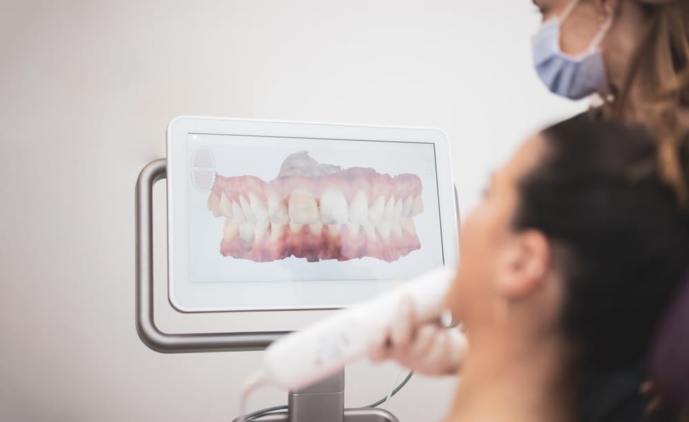
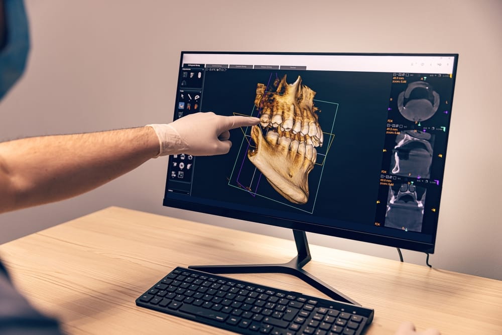
Cone-beam computed tomography, or CBCT, gives the dental team a three-dimensional view of the jaw before any procedure starts. A CBCT scan maps bone deficiencies and marks the edges of the defect. The scan also helps the team choose the right augmentation approach. CT scan data also guides implant positioning against available bone volume, removing the guesswork.
After surgery, follow-up imaging confirms that bone formation is progressing as expected. Implant placement only moves forward once that imaging shows adequate volume. Patients receiving full mouth dental implants benefit from this same imaging process throughout treatment.
Local anesthesia and local anesthetics keep patients comfortable during surgery. Conscious anesthesia options are available for anyone who needs extra support through a longer procedure. Suture technique at closure depends on flap design and the membrane used. Post-operative instructions from the dental team guide recovery from that point forward.
Prosthetic rehabilitation is the actual end goal of this whole process. It means placing titanium implants and the restoration above them. Bone atrophy and dental trauma both create the conditions that make GBR necessary. The result is a real path back to a functional bite and natural facial structure.
Ready to talk bone grafting for dental implants?
Bone grafting for dental implants gives your jaw the foundation it needs when bone volume falls short on its own. Guided bone regeneration combines barrier membranes, graft material, and CBCT imaging to build that foundation safely and predictably. The simultaneous or staged approach depends entirely on how much bone loss is present at your specific site.
If you’ve been told you don’t have enough bone for an implant, that’s a common starting point. Gum disease and other oral health issues can also play a role. Most patients are candidates once the dental team properly maps the defect. Schedule a consultation with us to find out which path fits your smile.
FAQs
How should patients care for the surgical site during healing?
Follow the post-operative instructions from your dental team closely. Most dentists recommend gentle rinsing for the first few days. Avoid brushing near the site early in healing. A modified hygiene routine protects the membrane and lowers the risk of wound dehiscence.
Do dental implants hurt?
Most patients feel little to no pain during the actual procedure since local anesthesia (or sedation) numbs the area completely. Mild soreness, swelling, or tenderness for a few days afterward is normal and manageable with over-the-counter pain relievers. Many patients say the recovery is easier than they expected, especially compared to a tooth extraction.
What is bone grafting?
Bone grafting is a procedure that rebuilds jawbone volume in areas where bone has thinned or been lost, often from missing teeth or gum disease. It uses graft material, sometimes from your own body, to support new bone growth. This creates a stronger foundation so a dental implant can be placed securely.
How long does it take for dental implants to heal?
Healing time varies by patient, but implants typically need several months to fuse with the jawbone through a process called osseointegration fully. Simple cases may move faster, while bone grafting or more complex sites can take longer. Your dentist will confirm readiness with imaging before placing the final restoration.
Is dental implant surgery painful?
Surgery itself isn’t painful thanks to anesthesia, and many patients describe only mild discomfort afterward, similar to a tooth extraction. Swelling, tenderness, or slight bruising can last a few days and usually responds well to standard pain medication. Sedation options are also available for patients who feel anxious about the procedure.