
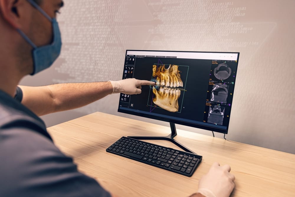
Planning for whole mouth dental implants starts long before surgery day. The anatomy beneath the gums decides whether a procedure goes smoothly or runs into trouble. Bone density, nerve pathways, sinus cavities, and blood vessels all affect where implants can safely go. Cone-Beam Computed Tomography imaging and digital surgical guides give the team a precise anatomical picture before any incision is made. This planning stage has changed how surgeons map and place full-arch implant cases.
Key Takeaways
- CBCT imaging shows bone, nerves, and sinuses in 3D, detail that flat X-rays cannot capture.
- Digital planning software turns CBCT data into a custom surgical guide built for one patient.
- Guided surgery can lower placement errors in full-arch and All-on-4 procedures.
- Pairing CBCT imaging with guided surgery supports safer, more predictable outcomes.
- Quick answers to common questions about CBCT imaging and full-arch implants are below.
What 3D CBCT Imaging Reveals That Dental X-Rays Cannot
Cone-Beam Computed Tomography, or CBCT, has become the standard of care in dental implant planning. It gives clinicians something traditional dental X-rays cannot: a full three-dimensional picture of the jaw, since conventional imaging is unable to depict complicated three-dimensional anatomical structures, as noted in BMC Oral Health. Standard dental X-rays produce flat, two-dimensional images that compress depth into a single plane.
What a Flat X-Ray Leaves Out
That flat view hides several details that matter most before implant surgery:
- How much usable bone is actually available
- Where the sinus cavities sit in the upper jaw
- How close nerve pathways run to planned implant sites
None of that shows up clearly on a standard X-ray.
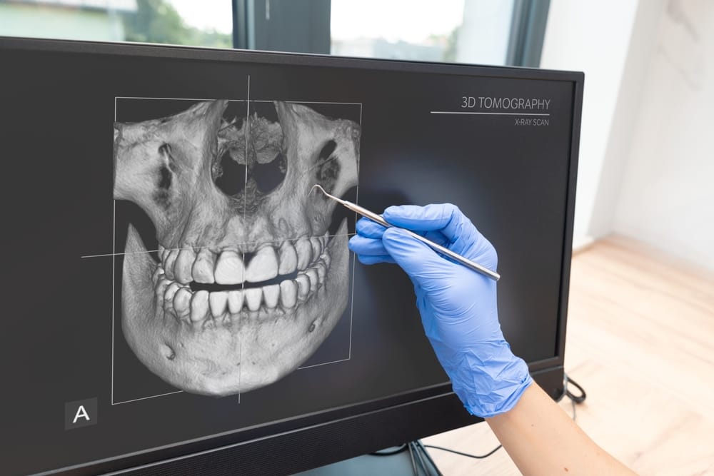
A CBCT scan captures all of it in a single pass. The scanner rotates around the head and produces a cone-shaped beam of X-rays. That beam builds a layered 3D model of the jaw. The resulting scan shows bone density, bone structure, blood vessels, and the exact path of the inferior alveolar nerve. This is the nerve most at risk during implant placement. The CBCT scan maps sinus locations in the upper jaw with the same level of detail.
How CBCT Data Supports Safer Placement
Research from the NIH National Library of Medicine indicates that CBCT lets clinicians assess the quantity and quality of the remaining bone. It also helps evaluate sinus position and map nerve pathways before surgery begins. Those measurements separate a safe procedure from a risky one. Nerve damage and sinus intrusion are the two most serious complications in implant surgery.
Jawbone density assessment through CBCT also flags bone defects that could affect implant stability. If bone has thinned in certain areas, that shapes the treatment plan before surgery starts, not during it.
Cone-beam imaging can also help the team evaluate the temporomandibular joint. It can spot craniofacial abnormalities that might affect how implants sit and function. When bone loss is significant, this same data helps determine whether bone grafting is needed first. Patients then know their full timeline in advance, rather than finding out mid-procedure.
Does a CBCT scan expose patients to high radiation levels?
Radiation from a CBCT scan is much lower than a medical CT scan. The dose is close to what patients receive from a full-mouth series of conventional dental X-rays. The imaging accuracy from a 3D CBCT imaging system outweighs that small amount of exposure. No other imaging option currently available shows this level of anatomical detail before implant surgery.
How Digital Surgical Guides Are Built From CBCT Data
The CBCT scan is the foundation. The surgical guide is what turns that data into physical precision during the procedure.
From CBCT Scan to Virtual Patient
Once digital imaging is complete, the dental team imports the scans into digital planning software. This software builds a virtual patient, a full digital model of the jaw, bone, and soft tissue. That model is based on scan data shown to deliver high accuracy and reliability for bony linear measurements.
Using computer-based planning tools, the team maps every implant position in three dimensions before surgery begins. This process generally follows a set sequence:
- Import the CBCT scan into planning software
- Test implant angle, depth, and path through virtual simulation
- Align the digital plan with the patient’s actual anatomy
- Generate a digital surgical guide from the finalized plan
- Produce the physical guide through 3D printing
The physical guide, sometimes called a stereolithographic surgical guide or surgical template, fits over the teeth or gum tissue. It contains precisely positioned channels that direct the drill to the exact location and angle set in the digital plan. This removes much of the variability that comes with freehand technique and replaces it with a controlled, repeatable process.
Why Guided Surgery Improves Accuracy
A randomized controlled trial published in PMC compared fully guided implant surgery with freehand placement. It found better placement accuracy, shorter surgical times, and fewer complications with the guided approach. Patient satisfaction scores in that study reached 9.2 out of 10. That reflects what guided surgery aims to deliver: consistent results across patients, regardless of how complex the anatomy is.
Clinicians measure implant placement accuracy in guided surgery by the gap between planned and actual position after placement. Custom guides produced through CAD/CAM systems consistently reduce that gap compared to freehand technique. For patients, that means a better restoration fit, less time in surgery, and lower risk near nerves or sinuses.
Digital registration, the process of aligning the guide with a patient’s real anatomy, is a key factor in final accuracy. When the digital model closely matches the physical jaw, computer-guided surgery removes much of the guesswork. This gives the surgical team a reliable reference point throughout the procedure.
All-on-4 Dental Implants and the Clinical Case for Guided Surgery
Full-arch cases raise the stakes in implant surgery. Replacing an entire arch of missing teeth means placing multiple implants at coordinated angles. Any shift from the planned position can affect how the final restoration fits and how load spreads across the jaw. It can also affect whether the implants reach long-term stability. This is where CBCT data and digital surgical guides make the biggest clinical difference.
How All-on-4 Implants Use Angled Placement
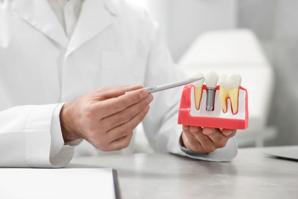
All-on-4 implants use four implants placed at specific angles to support a full prosthetic arch. Clinicians angle the back implants to use available bone and avoid the sinus cavities in the upper jaw. For patients with significant upper-jaw bone loss, sometimes called maxillary atrophic bone, this level of planning is not optional. Without it, the risk of sinus intrusion or implant failure increases.
A 2024 systematic review of guided implant surgery in fully edentulous patients examined static surgical guides made from CBCT data. It confirmed these guides consistently reduce the gap between the digitally planned and actual implant positions. That reduction affects implant stability, how well the implant bonds with bone, and how accurately the final restoration fits. The team identifies bone defects that could complicate surgery and builds them into the digital plan before the patient arrives.
Full-arch restorations also place prosthetic materials under higher functional loads than single-tooth replacements. Getting implant position right from the start reduces stress on the prosthetic components over time. Guided placement, backed by CBCT data, gives the restorative team the positional accuracy needed to design a prosthetic arch. That arch spreads force evenly across all four implants.
Where Implant Technology Is Headed
Implant technology keeps advancing. Researchers are studying robot-assisted placement and robotic-assisted surgery systems. They are also exploring virtual reality applications that let surgical teams rehearse procedures before operating on a patient. Clinicians are also applying artificial intelligence to CBCT data to help flag potential complications during the planning phase. These tools are not yet standard in most practices, but they show where digital dentistry is heading.
For patients considering full-arch restoration, it helps to know what the planning process actually involves. Understanding what a scan shows and how the team builds a guide gives a clearer picture of what to expect. It also explains why each step in the process exists.

Why CBCT Planning Makes Whole Mouth Dental Implants Predictable
Whole mouth dental implants depend on the planning that happens before surgery, not just the procedure itself. CBCT imaging gives the surgical team a three-dimensional map of the jaw, nerves, and sinuses well before the first incision.
Digital surgical guides then carry that plan into the operating room with a level of accuracy freehand technique cannot match. For full-arch restoration patients, these two tools reduce risk, improve accuracy, and give the final restoration a better fit. Wondering how this applies to your own anatomy and goals? Visit Dental Design Studios today!
FAQs
Is CBCT imaging safe, and how much radiation does it involve?
CBCT imaging uses a radiation dose close to what patients receive from a full-mouth series of conventional dental X-rays. It is much lower than a medical CT scan, and most clinicians consider it a proportionate diagnostic tool for implant planning, given the detail it provides before surgery.
Are dental implants safe?
Yes, dental implants are considered a safe and well-established treatment when placed by a trained professional. As with any surgical procedure, there are risks like infection or nerve irritation, but these are uncommon. Most patients experience a smooth recovery, and implants have a strong long-term track record for both safety and function.
What are dental implants made of?
Most dental implant posts are made of titanium, a metal known for bonding well with bone and fitting comfortably in the body. Some patients choose zirconia implants instead, which are metal-free and tooth-colored. The visible crown on top is typically made from porcelain or a similar durable, natural-looking material.
How long does an implant last?
Dental implants are built to last many years, and with good oral hygiene and regular checkups, the implant post itself can often last a lifetime. The crown on top may need replacement after 10 to 15 years due to normal wear. Diet, oral care habits, and overall health all play a role in longevity.
How long does a dental implant take?
The full implant process typically spans several months from start to finish, though the timeline varies by patient. Placing the implant post is often a single appointment, but healing and bone integration can take 3 to 6 months before the final crown is attached. Some cases allow for faster, same-day options.